i-CAT Technology
Advanced Imaging
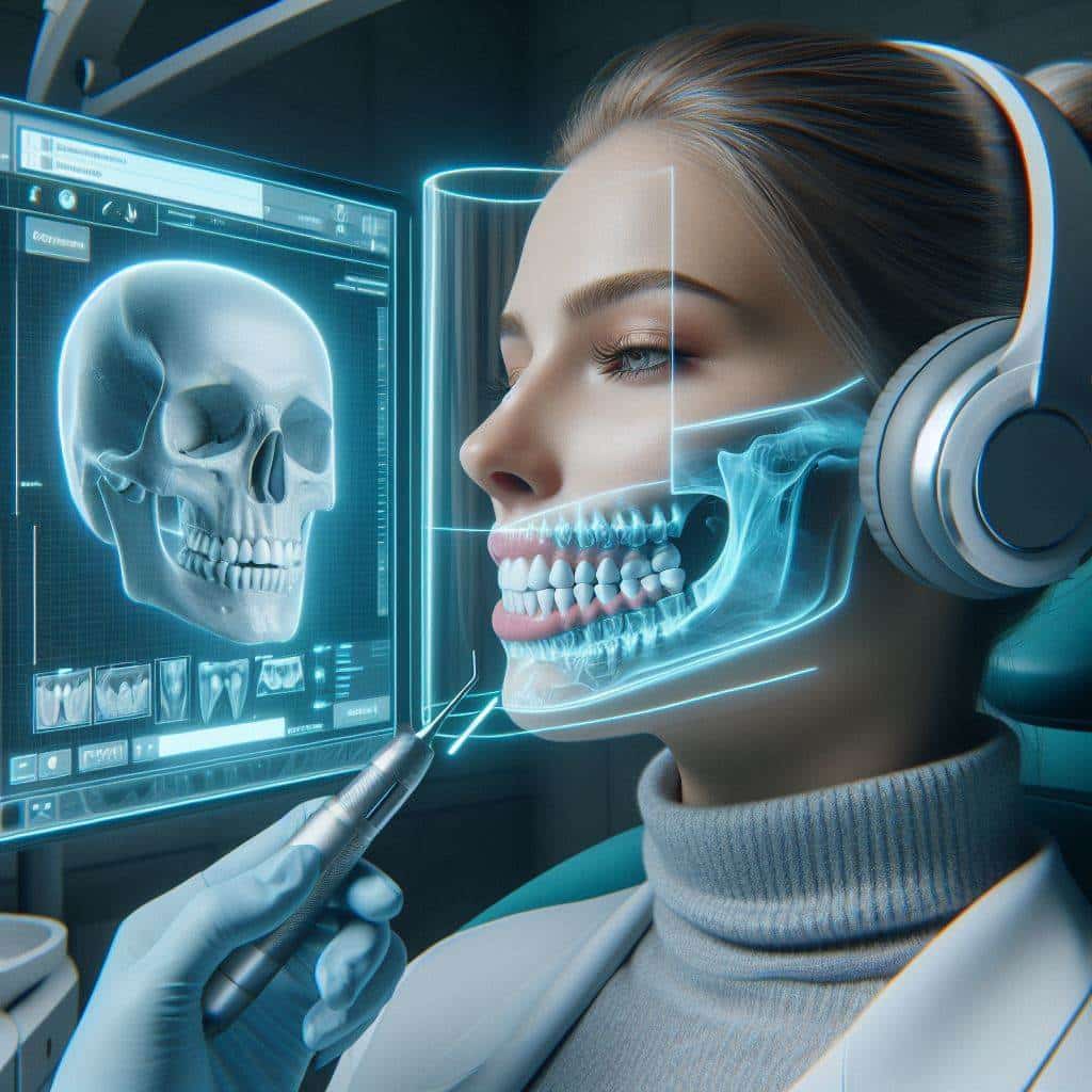
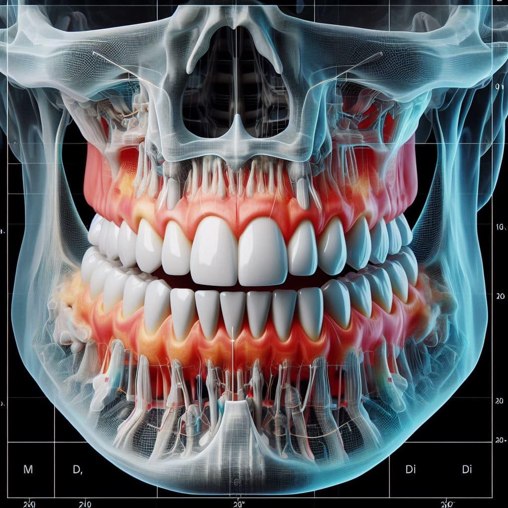
i-CAT technology provides high-resolution 3D dental imaging, offering detailed views of teeth, bones, nerves, and surrounding structures.
Minimally Invasive Treatment
With detailed imaging, i-CAT helps dentists plan minimally invasive procedures like dental implants, root canals, and orthodontic treatments…
Improved Patient Communication
Dentists can use i-CAT images to visually explain dental conditions and treatment options to patients, enhancing communication and understanding.
Precise Diagnosis
It enables dentists to accurately diagnose dental issues such as impacted teeth, bone abnormalities, and TMJ disorders, facilitating more effective treatment planning.
Customized Treatment
i-CAT allows for precise customization of treatment plans tailored to each patient’s unique dental anatomy and needs, optimizing treatment outcomes.
Enhanced Safety
i-CAT technology uses low radiation doses and fast scan times, prioritizing patient safety while delivering comprehensive diagnostic information for dental care.
I-CAT Technology Dental
Proudly Serving The Greater San Diego Area
Presently Dr. Korel now uses advanced 3D treatment tools for implants and restorations, including oral and maxillofacial surgery, TMJ and sinuses, and orthodontics. We aim to contribute to the continual improvement towards dental treatment. This why we are excited to have direct access to i-CAT® scanners, the best ever cone beam scanner.
The i-CAT scan generates a high definition 3D diagnostic images for greater treatment effectiveness. You can now get a comprehensive view of the 3D models of your jaw and bite in our office due to the new i-CAT scans that are available. This implies that Dr. Korel and his team can correctly capture the different anatomy of each patient and improve therapy progress. The flexibility of i-CAT enables to obtain the most exceptional advantage to each patient’s treatment plan, from diagnosis, through planning and treatment, to successful case completion.
About I-CAT™ 3D Dental Imaging Technology Services
The I-CAT™ Cone Beam 3-D Dental Imaging System produces high-definition, in-office, 3D, digital imaging for higher performance and increased accuracy at a reduced price, as well exposes patient to less radiation than traditional CT scans. It provides faster and more accessible image caption. Implants and oral surgical operation are concluded within minutes as the i-CAT scan allows analysis of delicate anatomy and appraisement. It collects more anatomical data about the structure of the bone and tooth alignment for the usual type of implant to choose treatment and placement. I-CAT identifies and points out problems before they become critical by maintaining accuracy while measuring bone and jaw deformities, determining bone lesions and the jaw and detecting other pathologies that may be infectious and cause severe tumors and disease. Several projections are needed to determine tooth relationship correctly and sufficiently sustains the actual representation of anatomy provided as this improves implant diagnosis and treatment. I-cat provides the wide view of the position of the tooth and association of the abnormal anatomy with a more accurate viewing of impacted supernumerary or abnormal teeth in association with other anatomical structures, including the roots, nasal fossa and sinuses to improve correct management of treatment by knowing the tooth’s position and its relationship to adjacent teeth and structures. Since the concentration of this imaging is a narrow part of the body, other medical specialists may need imaging if your situation requires it.

BENEFITS OF I-CAT
The i-CAT scanner has a various number of distinct advantages over traditional impressions, including:
- The scanning process is fast & convenient.
- Provides an accurate three dimensional model of the teeth and jaws.
- Enables to manipulate the model digitally.
- I-CAT dental makes it easier to plan a personalized therapy.
- Exact measurements of relevant anatomical factors are created.
- It also enhances accuracy and treatment effectiveness.
- 3-D Imaging and Dental Reconstruction
Dental reconstruction will be easier to plan, and high level of accuracy is attained during placement with the use of these 3-D images. TheiCat dental will display dimensions and angles that dentists require to be aware of, which leads to accurate placement reconstruction. Also, the image can display the density of the bone so that it is well noted before the reconstruction work commences. Cutting down is well planned on the amount of time required, once all of this information can be collected immediately. During one sitting the dentist will have to check the images that produced and think of a plan best suitable for that patient’s individual requirements before they must have left the office after their first appointment.
i-CAT Technology Can Assist with TMJ & Sleep Apnea
TMJ
Images can serve as a medium to concentrate on a particular area of a patient’s body structure, including the temporomandibular joint. Diagnosing situations like TMJ is considered easy with the use of images captured by the i-CAT. We can assess more accurately the amount of misalignment that a patient with TMJ has in order to derive a therapy plan following this technology. I-CAT scan has the ability to find the areas where teeth do not line up appropriately which may lead to the cause of TMJ since the images caption the entire mouth.
SLEEP APNEA
We will be able to detect blockage of airways that may lead to sleep apnea with the use of this technology. To determine the suitable therapy options the amount of inflow of air has to be determined. To figure out whether it is or is not sleep apnea symptoms that are related to more than just restrictions in the airway itself, the structure of the mouth can also be analyzed Other breathing difficulties which include abnormalities in the sinuses will even show up in iCat scans.

Choose Accuracy & Speed
Selecting a dentist who uses this new and efficient technology could enhance your experience also as well as the care you receive. Each patient mouth is different and should be necessarily treated according to your specific needs. By Investing in this I-CAT™ Cone Beam 3-D Dental Imaging System enables us to better understand your unique needs so we can perform more authentic care.
Utilizing i-CAT Technology: Dr. Korel now has direct access to advanced 3D treatment tools for implants and restorations, oral and maxillofacial surgery, TMJ and sinuses, and orthodontics.
i-CAT produces high definition 3D diagnostic images for ultimate treatment efficiency. This means that Dr. Korel and his team can accurately capture each patient’s unique anatomy and treatment progress.From diagnosis, through planning and treatment, to successful case completion, i-CAT’s flexibility allows Dr. Korel to gain the greatest benefits to each patient’s treatment plan.
CONTACT US TODAY FOR THE BEST CARE
We are excited to announce the availability of the i-CAT scanner in our office and have a great impact on our commitment to the well-being and convenience of our estimated patients.
WITH YOUR DOCTOR
Suspendisse venenatis erat et sem egestas, sit amet luctus urna aliquet. Suspendisse cursus efficitur justo. Nam pulvinar sagittis lacus, varius scelerisque nisl pellentesque nec.
Select your preferred service and book a dentist visit in minutes. We’ll remind you of your appointment by SMS
WORKING HOURS
CONTACT DETAILS

At Korel Dentistry Implant & Cosmetic Dentist in San Diego, CA we believe a beautiful smile reflects your well-being and confidence. Our dedicated team provides tailored care for your needs, ensuring a healthy and dream smile. From routine to cosmetic treatments, our state-of-the-art facilities and compassionate approach guarantee outstanding results. Schedule your appointment today for a brighter smile!
Website Links
The information on this website is for general information purposes only. Nothing on this site should be taken as medical advice for any individual case or situation. This information is not intended to create, and receipt or viewing does not constitute, a doctor-patient relationship. Website Crafted with Love and Coffee by SandiWeb® : Web Development and Marketing Solutions. ©1998 – 2024. Korel Dentistry Implant & Cosmetic Dentist All Rights Reserved.

